Ultrasound Price in Ranchi
The ultrasound price in Ranchi can vary depending on the type of ultrasound, the diagnostic center, and the complexity of the procedure. On average, a routine abdominal ultrasound can range from ₹800 to ₹2,500, while more specialized ultrasounds like obstetric or cardiac scans may cost anywhere from ₹2,500 to ₹5,000 or more.
It is advisable to contact us in Ranchi to get accurate pricing details. Ensure you choose us for accurate results and a positive experience.
Ultrasound Test in Ranchi
Ultrasound, also known as sonography, is a widely used diagnostic tool that helps medical professionals view and assess internal organs, tissues, and blood flow. It uses high-frequency sound waves to produce images, making it non-invasive and highly effective for diagnosing a wide range of health conditions. In this article, we’ll cover everything you need to know about ultrasound tests, including the procedure, costs, preparation, and types of conditions detected through ultrasound.
Alternative Names for Ultrasound
Ultrasound is often referred to by several names. Commonly, it is also called:
- USG (Ultrasonography)
- Sonography
- Echo (when used to assess the heart, e.g., Echocardiogram)
- Ultrasonic Imaging
Despite the different names, the procedure remains the same, and it’s used to diagnose various medical conditions.
Preparation for Ultrasound
Preparation for an ultrasound largely depends on the type of examination being conducted. Some general tips for preparing for an ultrasound include:
-
Fasting: For abdominal ultrasounds, you may be asked to fast for a few hours before the test. This helps get clearer images of your organs like the liver, kidneys, and pancreas.
-
Hydration: For pelvic ultrasounds or obstetric ultrasounds, you may be asked to drink plenty of water before your appointment, as a full bladder can help provide better images of the pelvic region.
-
Clothing: Wear comfortable clothing that allows easy access to the area being examined. It’s often recommended to wear loose-fitting clothes so the technician can easily apply the ultrasound gel and use the transducer on your body.
-
Other Instructions: Follow any specific instructions provided by your healthcare provider regarding your ultrasound.
What Happens During the Ultrasound Test?
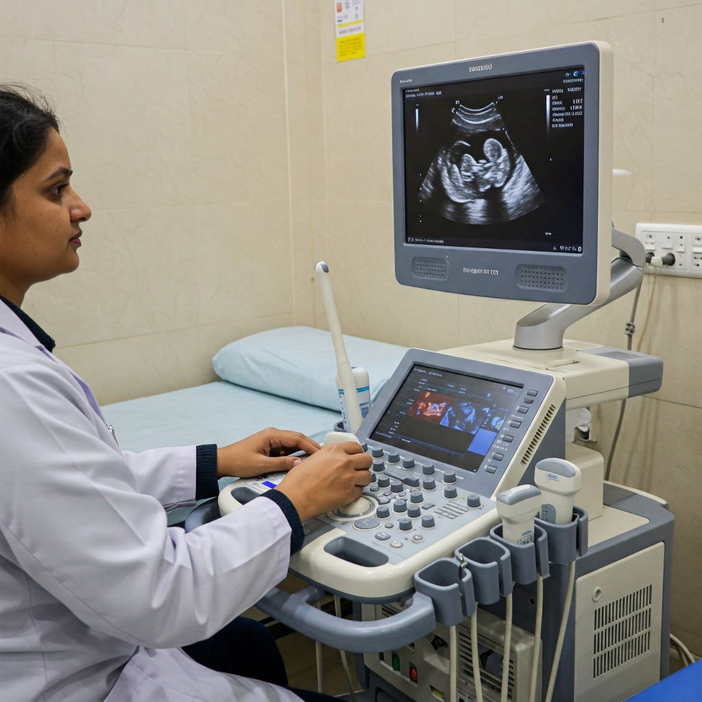
During the ultrasound, a gel is applied to the area being examined. This gel helps the sound waves travel more effectively and improves the quality of the images. The technician or sonographer will use a device called a transducer, which looks like a small wand, to move over the gel-coated area. The transducer emits sound waves, which bounce off the tissues and organs inside the body, and the echoes are converted into images that are displayed on a monitor.
For some ultrasounds, you might need to lie on your back or side, while for others, you may be asked to change positions to get better images.
Is an Ultrasound Painful?
No, an ultrasound is not painful. Most people experience little to no discomfort during the procedure. The transducer may apply some pressure to the area being examined, but this is generally mild. The gel can feel cool and slightly messy, but it is completely safe. If you’re undergoing a transvaginal ultrasound, the procedure might involve the insertion of a probe into the vagina, which some individuals find uncomfortable but not painful.
Are Ultrasounds Safe?
Ultrasounds are considered very safe. Unlike X-rays or CT scans, ultrasounds do not use ionizing radiation, which makes them a safer choice, especially for pregnant women. Since ultrasound uses sound waves, it is non-invasive and does not pose any known long-term risks to the body. It is commonly used during pregnancy to monitor fetal development, and its safety profile makes it suitable for various other medical conditions as well.
What Are the Conditions Detected by Ultrasound?
Ultrasound is an incredibly versatile diagnostic tool, capable of detecting various health conditions. Some of the most common conditions detected by ultrasound include:
-
Pregnancy Monitoring: Ultrasound is widely used to monitor the development of the fetus, detect multiple pregnancies, and identify issues such as ectopic pregnancies or fetal anomalies.
-
Gallstones: Abdominal ultrasound can identify the presence of gallstones in the gallbladder, helping diagnose related conditions like cholecystitis.
-
Kidney Stones: Ultrasound is often used to detect kidney stones and monitor their size and position.
-
Liver Disease: Liver conditions such as cirrhosis, fatty liver disease, or tumors can be identified using ultrasound.
-
Heart Conditions: Echocardiography, a type of ultrasound, is used to evaluate the heart’s structure and function, detecting issues like valve problems, heart failure, or congenital heart defects.
-
Cysts or Tumors: Ultrasound can help detect cysts or tumors in organs like the ovaries, thyroid, and breasts.
-
Blood Clots and Vascular Problems: Doppler ultrasound helps assess blood flow and can detect conditions like deep vein thrombosis (DVT) or arterial blockages.
-
Musculoskeletal Injuries: Soft tissue injuries, tears in ligaments or tendons, and joint issues can be examined using ultrasound.
Types of Ultrasound
There are several different types of ultrasound tests, each used for specific purposes:
-
Abdominal Ultrasound: Used to examine the organs in the abdomen, such as the liver, kidneys, pancreas, and spleen.
-
Pelvic Ultrasound: Focuses on the organs in the pelvic area, including the uterus, ovaries, and bladder.
-
Obstetric Ultrasound: Primarily used to monitor pregnancy, assess fetal development, and detect any potential complications during pregnancy.
-
Doppler Ultrasound: Measures blood flow through blood vessels, helping detect issues like blood clots or narrowing of arteries.
-
Echocardiogram (Heart Ultrasound): An ultrasound used to assess the heart’s function and structure, identifying conditions such as heart valve disease or heart failure.
-
Transvaginal Ultrasound: Used to get detailed images of the female reproductive organs by inserting a probe into the vagina, often used for early pregnancy monitoring or checking for ovarian cysts.
-
Musculoskeletal Ultrasound: Used to assess soft tissues, joints, muscles, and tendons for injury or disease.
Ultrasound Result
Once the ultrasound procedure is completed, the images captured are reviewed by a trained radiologist or the attending physician, who interprets the findings. Results are typically available within a few hours to a day, depending on the diagnostic center. In some cases, the results may require further testing or consultation with a specialist.
Your healthcare provider will explain the ultrasound results and discuss any necessary next steps, such as treatment or follow-up tests, depending on what was detected.
Conclusion – Ultrasound Price in Ranchi
Ultrasound is a safe, non-invasive, and versatile diagnostic tool that plays a crucial role in detecting a variety of medical conditions. Whether you’re expecting a baby, checking for internal organ issues, or assessing joint and muscle health, ultrasound provides accurate and reliable results. If you’re looking for ultrasound tests in Ranchi, ensure you choose a diagnostic center with experienced professionals and advanced technology for the best care.
Also, contact us for CT Scan in Ranchi